

I am sure you must have searched.
How to care for the skin?
Vitamins for glossy skin.
Best skincare product. Etc
But have you searched “Structure and most important functions of the skin”? Let me explain to you “Structure and 11 most important functions of the skin“.
So start with” what is skin?”
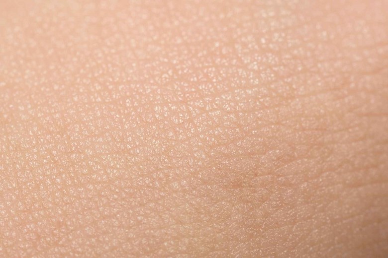
Skin is a part of the integumentary system which is a natural protective outer body covering of humans or animals.
What is the integumentary system?
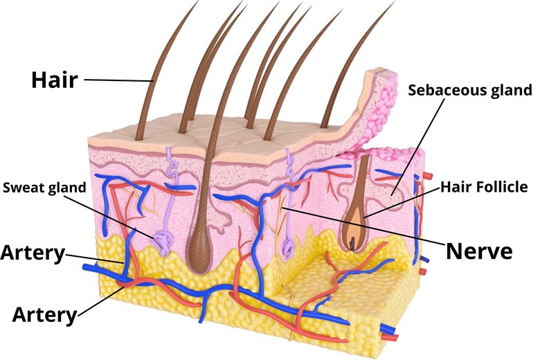
The skin and its appendages (hairs, nails, glands, nerves, etc) are called the integumentary system.
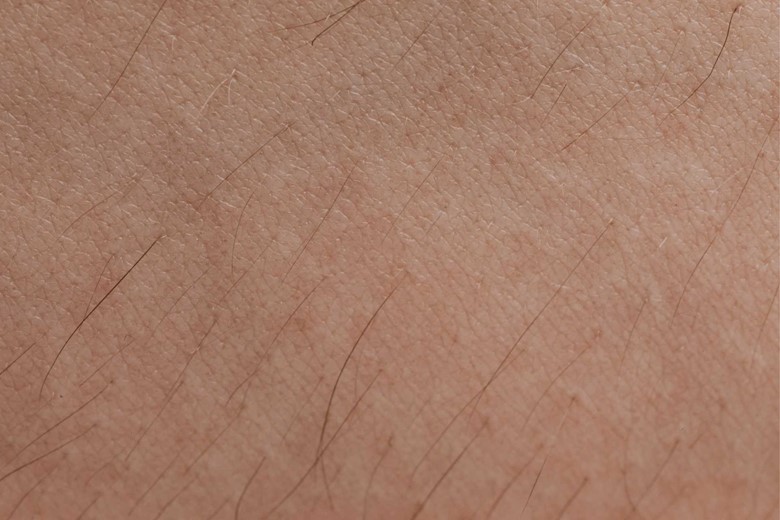
Skin covers all the bodies of a person and is the largest organ of the body.
Is their thickness the same all over the body?
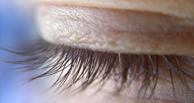

No, it varies in thickness. On the eyelid, it is 0.5mm thick while on the palm of hands and sole of feet it is 4mm thick.
Why the eyelid has minimum thickness while the palms and soles have maximum thickness?
Skin is made of different layers which will be explained later here. Eyelids have no subcutaneous layer which makes them thin from other parts skin.
While palms and soles have a maximum thickness to withstand high pressure and physical stress but remember, research shows that thickness beyond normal is a sign of esophagus cancer (Tylosis) which causes the severe thickness of palms and soles.
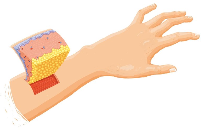
Can you explain the structure of the skin?
Skin is made of primary two layers.
- Epidermis
- Dermis
- Hypodermis (not a part of true skin)
1. Epidermis
“Epi” word comes from the Greek language which means “over” or “upon” so it means epidermis is the outermost layer of skin.
It is a waterproof and protective barrier over the body which serves against infections.
The epidermis is further divided into different layers. They are:
a) Basement membrane
The basement membrane separates the epidermis from the dermis below. It is made up of cuboidal epithelial cells and forms a layer called stratum Basale.
What do these cells or layers do?
Well, these cells (stratum Basale) divide, and as these cells divide they move up and lose the blood supply and become flattened which are then called keratinocytes.
Stratum Basale has a type of cell called MERKEL CELLS which play important role in sensation and are thought their function as the senses of light touch, and hair movement. Some of these cells are also found in the dermis.
b) Stratum spinosum
As stratum Basale cells divide they migrate above and form another layer called stratum spinosum.
Types of cells in Stratum Spinosum
i) Melanocytes
Stratum Spinosum has melanocytes which are protein pigments (melanin) producing cells. These cells are also found at the base of hair follicles where it transfers the melanin to keratinocytes which make up hair.
What are the functions of melanocytes?
Pigmentation
Melanocytes are responsible for pigmentation by producing melanin (pigments) in different people of the world and their pigmentation range from black to reddish-yellow. How much these pigments are metabolized gives color to different people.
Protection from UV light
When keratinocytes are exposed to the sun they send chemical signals to melanocytes to produce melanin. The melanocytes then produce melanin and transport it in small sacs called melanosomes to keratinocytes where it is metabolized and functions as a natural sunscreen and scatters the UVB light which destroys the skin DNA and can cause skin cancer if exposed without it.
ii) Langerhans cells
It is a type of dendritic cell. These cells are antigen-producing cells that play important role in immunity.
c) Stratum Granulosum
When Stratum spinosum moves up and forms another thin granular layer called stratum granulosum.
What is the function of this layer?
These cells contain keratohyalin granules which are filled with histidine- and cysteine-rich proteins that serve to bind keratin together.
d) Stratum Lucidum
Above the stratum granulosum is a thin, clear dead cells layer called stratum lucidum which is only found in a thick area of the body i.e palms of hands and soles of feet.
e) Stratum Corneum
It is the outermost layer of the epidermis and maybe the thickest layer of the epidermis.
What is their function?
They have a protective and adaptive physiological function like protection of underlying tissues from infection, dehydration, chemical, mechanical stresses, microbial proliferation, initiation of inflammation, etc.
Can you tell, which layer of skin is shed regularly?
Stratum corneum layer cells are regularly shed and that process is called Desquamation. The shedding process regulates the formation of new keratinocytes and the migration from Stratum Basale to Stratum Corneum takes about 14 days.
2. Dermis
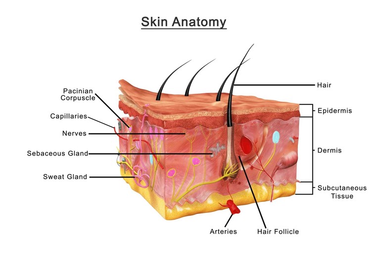
The dermis is the second and thickest layer of skin containing most of the structures. It is divided into two layers:
a) Papillary layer
The papillary layer is the top superficial layer of the dermis and is made up of loose connective tissues and thin collagen fibers.
Papillary layer form projections called papillary where it fits epidermal ridges of the epidermis and increase the surface area of these two layers.
Other structures found in the dermis
i) Capillaries
Capillaries originate in the dermis as well as in the hypodermis and move to the Papillary layer of the dermis where it connects and supplies nutrients to the Stratum Basale of the epidermis.
ii) Sensory Nerve
Papillary Layer also contains sensory nerve endings that receive a different stimulus.
b) Reticular layer
The reticular layer is the second layer of the dermis and is made of dense connective tissue and thick collagen fibers.
Why it is called the reticular layer?
Because it is made of thick collagen fibers connected with each other and forms a network, that’s why it is called the reticular layer.
Who makes this network of collagen?
There is a special type of cell called fibroblast which secretes and produces these collagen fibers.
Can you tell, what other structures are found in the dermis?
Yes, the following are the structures found in the dermis.
i) Hair Follicle
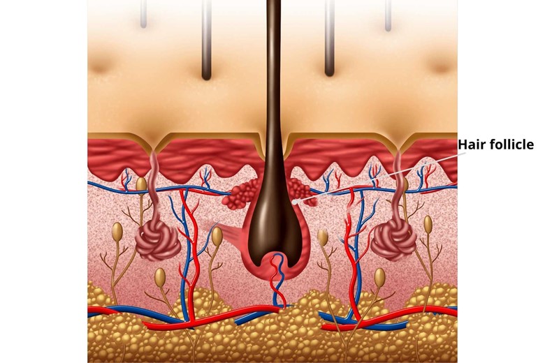
Hair Follicle is found in the dermis from where hair arises.
ii) Arrector Pili Muscle
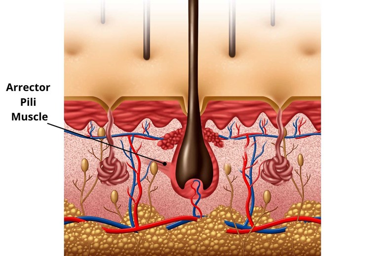
This is a tiny muscle that attaches to the base hair follicle. It contracts to erect the hair during goosebumps.
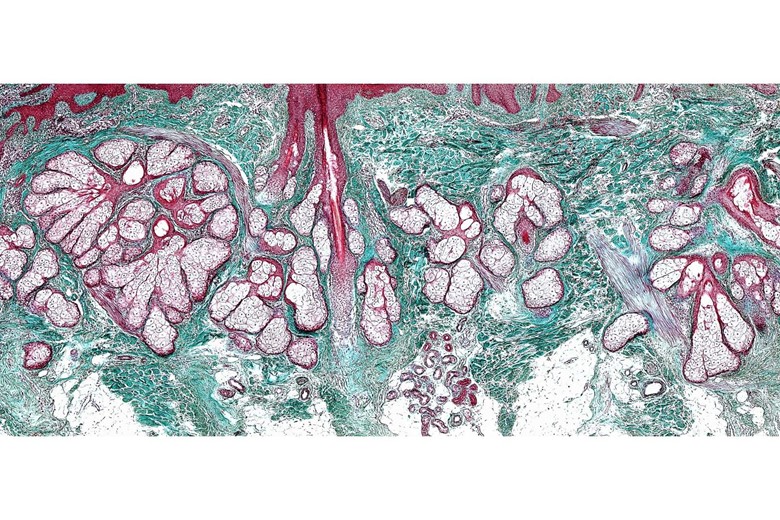
iii) Sebaceous glands
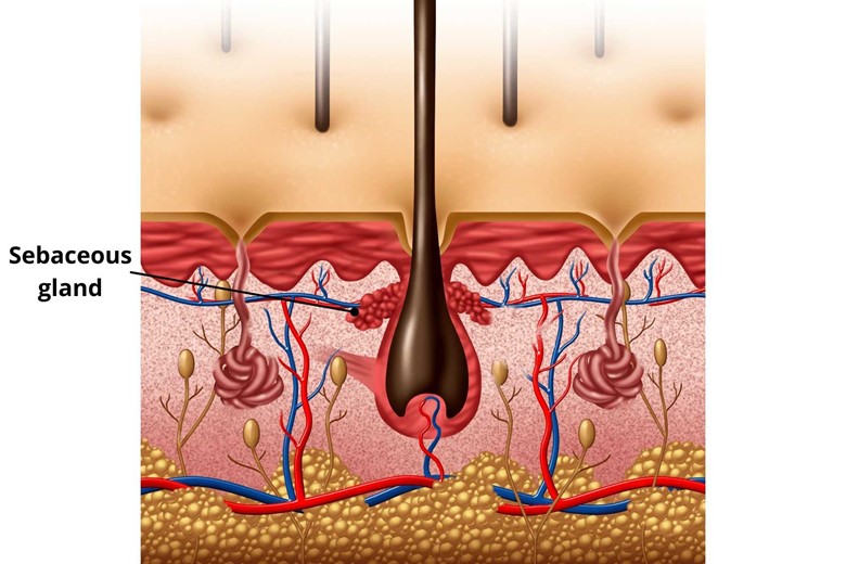
Sebaceous glands have two types, one connected to the hair follicle and the other which is independent.
The sebum (made of triglycerides, wax esters, squalene, and metabolites of fat-producing cells) produces a sebaceous gland whether it is present near to hair follicle or hairless area its function is to lubricate the hair and skin.
Sebum has also an important role in thermoregulation where it emulsifies sweat forming a sheet of sweat that is not lost in drops of sweat performing an important role in minimizing dehydration too.
iv) Apocrine sweat glands
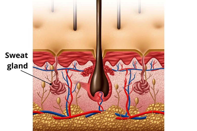
Apocrine glands are tubular and coiled structures and are found in hair-dense areas like axillae (armpits) perineum (the space between anus and scrotum) and genitalia. This gland is hormone-driven and is activated after puberty before puberty it is inactive.
v) Eccrine glands
Eccrine glands are found almost all over the body but with high density on the palm and soles than on the head but it is much reduced on the trunk and extremities. It is not present on lips, ear canal, prepuce, and genitalia.
It is ten times smaller than the apocrine gland and is not deeply extended in the dermis therefore they secrete its secretions directly on the surface of the skin.
Their secretion is called sweat which is mostly water and some electrolytes (sodium chloride) and functions as a tool down the body during high temperatures.
Skin color has an important role in their environment. People get their skin color according to their environment, let’s explain all these.
So what is the difference between white skin and black skin?

The difference is due to melanin (pigments). Both produce melanin which is called eumelanin and pheomelanin.
What are eumelanin and pheomelanin?
Eumelanin:

Eumelanin is pigments ranging from brown to black in color.
Pheomelanin:

It is produced in light-toned skin.
Why the black skin has more eumelanin?
Because eumelanin is a natural sunscreen protecting the skin from ultraviolet rays of sun which is the main cause of skin cancer. The eumelanin surrounds the nucleus when exposed to the sun and protects its DNA. As you know people are black in the warmer area of the world where sun rays are intense so the melanin is secreted in sufficient amounts to protect their skin from the dangerous effects of UV light.
Some people have eyes, hair, and skin all are white and vision defects, Is that a pigment?
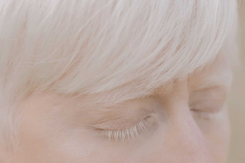
No, it is called albinism where there is a complete or partial absence of pigmentation in hairs, eyes, and skin. The person has many problems the most prominent is vision difficulty.
Albinism is a congenital and genetic disease that may receive from the mother or father after birth.
3. Hypodermis
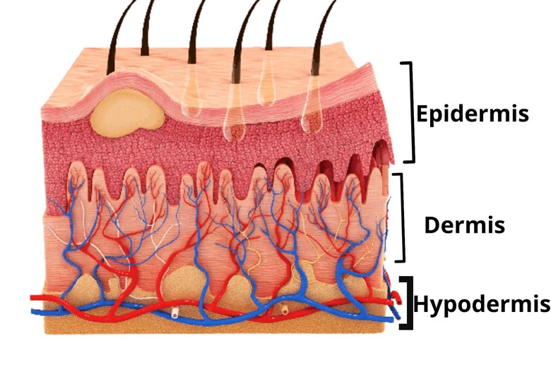
The hypodermis is derived from the Latin word meaning beneath the skin and it is not considered a part of true skin and is also called the subcutaneous layer, or superficial fascia. It is located between the dermis and underlying tissue and organs.
Hypodermis connects the skin to underlying fibrous tissue of muscle and bones.
What structures are found here?
Following are some of the structures found in the hypodermis.
a) Adipose tissue
Adipose tissue is nothing but just fatty cells that store energy and serve as an insulator for heat loss.
b) Areolar tissue
Areolar tissue is a loose connective tissue that binds the skin to the underlying muscles and provides strength, support, and elasticity. The loose connective tissue also provides a link between organs making a high degree of movement between adjacent body parts.
c) Other structures
The other structures found in the hypodermis are Macrophages, nerves, blood vessels, etc playing an important role in protection.
Can you tell the important functions of hypodermis?
i) Body temperature regulation
The adipose layer (fats) works as an insulator during winter when the body needs to conserve heat. In Whale which is an aquatic mammal, the blubber (fatty layer under the skin) serves as a major heat reservoir in the cold aquatic environment.
ii) Hormone production
Leptin hormone is secreted by fat cells of hypodermis which tells the body to stop eating.
We have discussed the first part i.e. Structure of our topic “Structure and 11 most important functions of the skin” Now let’s explain the second part of our topic.
We also called it the physiology of skin or functions of the skin.
Physiology of skin/ Functions of skin
1. Act as a Barrier
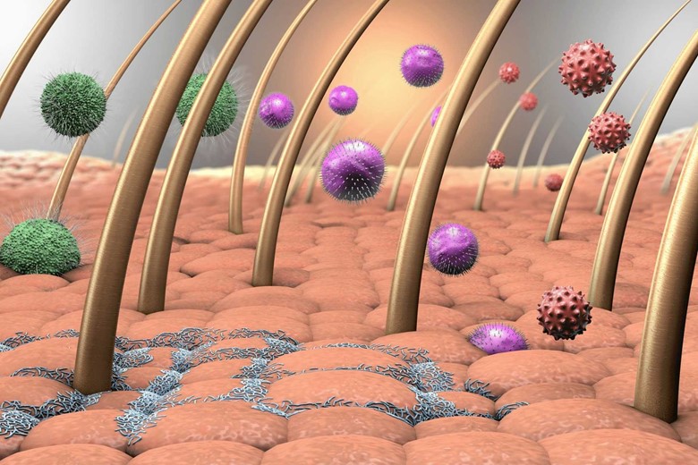
Skin act as a barrier for different agents. The skin has a lot of microorganisms (which we call normal flora) that are limited to the stratum corneum of the epidermis and are not allowed to enter the body thus protecting our body from their damage.
2. Protect the internal organs from damage
Skin also acts as a barrier against cuts and injuries. For example, during cuts or injuries, the skin minimizes damage to the underlying and inner tissues of the body.
3. Signal to immune response
Dendritic (Langerhans) cells in the skin respond to the immune system during injury or cut because there are bacteria on the skin if it is invaded it can cause more damage so dendritic cells signal to the immune system and the immune system responds to bacteria and protect the injury from further damage.
4. Protection from UV rays/cancer
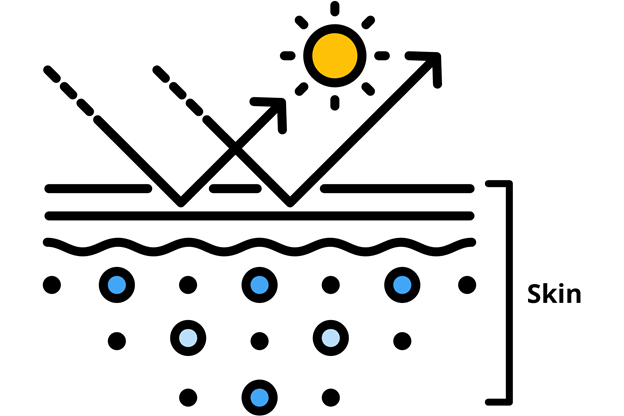
As already discussed the stratum spinosum has melanocytes which have melanin in small sacs called melanosomes. Upon exposure to sun rays, the melanocytes are activated which sends the melanosomes (melanin sacs) to keratinocytes (skin’s cells) and also to deeper tissues where the melanin surrounds the cell’s nucleus and protects their DNA from harmful effects of the sun, protecting skin from skin cancer.
5. Vitamin D synthesis
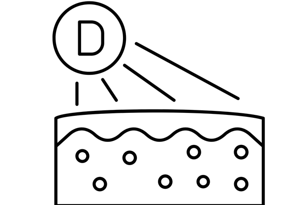
The skin has 7-dehydrocholesterol which is a zoosterol (sterols of animals are called zoosterol) and pro-vitamin (that can be converted to vitamin in the body). Upon exposure to the sun, the 7-dehydrocholesterol is converted to vitamin D3 (cholecalciferol). Vitamin D3 helps in the absorption of calcium and phosphorus in the small intestine making bone health good.
6) Temperature regulation
Skin regulates temperature by two methods.
i) Sweating
ii) Vasoconstriction and Vasodilation
i) Sweating
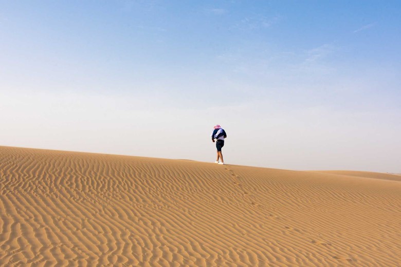
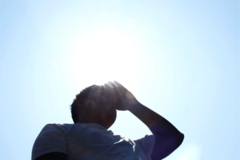
For example, you are in a desert or warmer area of Africa. And you know both are the hottest areas of the world and you also know their environment increases your body temperature.
Now what body will do?
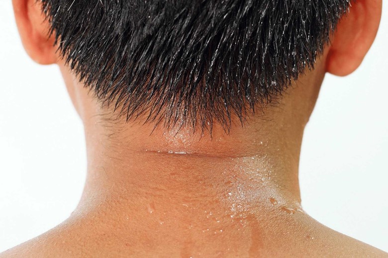
The hypothalamus of the brain detects an increase in temperature and sends stimulation signals to sweat glands to secrete more sweat (water, salt, and urea) producing a cooling effect because evaporation of sweat releases heat from the body and regulates the body temperature.
ii) Vasoconstriction and Vasodilations
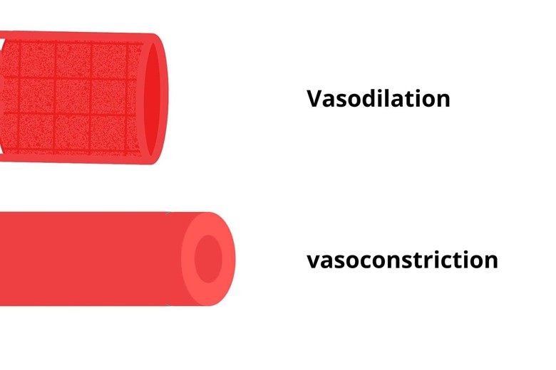
Vasoconstriction is the dilation of blood vessels in the dermis of the skin. Let’s take the same warm situation as above, now the hypothalamus signals the blood vessels in the skin (dermis) to dilate so that the blood flow to the skin increases causing the radiation of heat out of the body. Similarly, the blood vessels in the visceral (internal) body are contracted called vasoconstriction so that the blood flow moves from the internal body to the skin for radiation of heat.
Is this phenomenon the same in winter too?
No, in winter the phenomenon is the opposite i.e. the sweating is stopped, vessels in the skin contract (vasoconstriction), and the internal body vessels dilate (vasodilation). The vasoconstriction in the skin reduces the blood flow to the skin thus reducing the radiation of heat from the body as the body needs heat in the winter season while the vasodilation in the visceral body conserves the heat internally.
But during exercise or hard-working why does the skin become red?
The same phenomenon. The vasodilation in the skin and vasoconstriction in the visceral body thus making blood flow to the skin for radiation of heat which makes the skin red during the exercise.
7. Secretions
The skin secretes some secretions which are as follows.
i) Sebum
Sebum is released from sebum glands in the skin which lubricates the skin and hair.
ii) Sweat

Sweat contains water and electrolytes (e.g. sodium chloride) which are secreted from the skin thus playing an important role in water and electrolytes homeostasis.
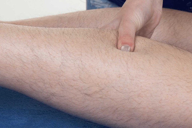
8. Provides strength and flexibility

Skin has collagen and elastic fibers. The collagen fibers provide strength to the skin while elastic fibers provide flexibility due to which the body moves in multiple directions without tearing the skin.
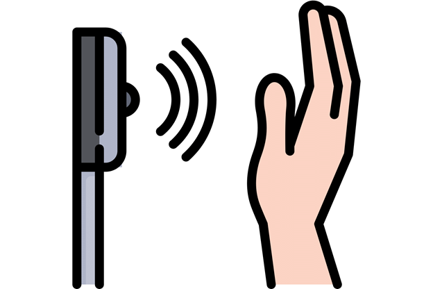
9. Sensory function
Following are the structure that performs the sensory function of the skin.
i) Meissner corpuscles/ touch receptor
Do you know the corpuscle?
No.
Okay, let me explain to you, a corpuscle is a “small body” or “minute body” or “cell” or “a particle” in the body.

Meissner corpuscle is also called tactile corpuscle, a type of mechanoreceptor (responds to mechanical pressure or distortion) that is responsible for sensitivity to light touch and low-frequency vibration.
Where this structure is located in the skin and on the body?
This is located in the upper dermis (dermis papilla) and below the epidermal ridges of the skin. On the body, it is mostly found in the fingers, lips, and toes.
ii) Pacinian corpuscles/ pressure receptor
Pacinian corpuscles are also called lamellar corpuscles because of their lamellated structure. It is located in the deep dermis and is responsible for the detection of fast vibration and pressure on the skin.
iii) Thermoreceptors
As the name indicates these receptors are for temperature detection. These receptors have free nerve endings.
What is meant by free nerve endings?
Free nerve endings mean they are not like corpuscular as we discussed in previous receptors like Meissner’s corpuscles and Pacinian corpuscle.
Again there are two types of thermoreceptors for detection i.e one is for cold temperature detection and the other is for hot temperature detection.
iv) Nociceptors/pain receptors
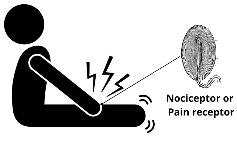
Nociceptors or pain receptors make the skin sensitive to pain. In injury i.e. cuts, burns, accidents, etc the Nociceptors/pain receptors receive the pain.
Do you know why burns are more damaging and panic than other injuries?
No, please explain.
when there is a burn or scratch on the road or stone or something hard, the upper epidermis blows away, and pain receptors are exposed to the environment causing more pain to the body than a cut.
v) Ruffini corpuscles
Ruffini corpuscles are also called Bulbous corpuscles or Ruffini ending and are responsible for skin stretch, contribute kinesthetic sense, and control finger position and movement.
10. Protection
Hypodermis has fat cells that not only function as storage of energy but also provide protection and serve as a cushion while sitting on hard structures like a chair, floor, and other hard sitting areas.
11. Shock absorber
The hypodermis serves as a shock absorber of the body protecting the body from shocks.
Conclusion/Summary:
The topic “Structure and 11 most important functions of the skin” show the importance of the structure of the skin because it is the first barrier that resists infections, accidents, burns, damage, etc, and most importantly it protects the DNA from the ultraviolet rays of the sun. Skin is primarily composed of two layers epidermis and dermis while hypodermis is not considered true skin but its borders are not clearly distinguished from the dermis that’s why sometimes it is also considered skin. And it is not just a single layer, it is composed of many layers internally (explained above) so that microorganisms don’t enter the internal body. The sebaceous glands in the dermis lubricate the skin and prevent the body from dehydration being fatty, the sweat glands regulate the body temperature both in a cold and hot environment.
In short, the whole discussion shows that our skin has very important functions for us so we need to take special care of our skin by providing nutrients and vitamins for glowing skin because it is the first line of defense and it became more important when there is any injury to the skin or any burn or scratch on a road or something hard thing is more painful than cutting because the pain receptors (nociceptors) after scratch or burn is exposed making it more painful. Are you caring for your skin tell in the comment box how you benefited?


2 thoughts on “Structure and 11 most important functions of the skin”